Best Retina Clinic in Chennai | Retina Specialists & Advanced Retina Treatment
Looking for the best retina doctor in Chennai? At Dr. Ashok Eye Hospital – Unit of Centre for Eye and Health Care, our experienced retina specialists in Chennai provide comprehensive diagnosis and advanced treatment for retinal diseases using the latest diagnostic technology and evidence-based care. Led by Dr. Ashok Rangarajan, our retina clinic offers expert care for diabetic retinopathy, retinal detachment, macular degeneration, retinal vein occlusion, retinal tears, macular hole, and other complex retinal disorders. With advanced OCT, OCT Angiography (OCTA), Fundus Imaging, FFA, Retina Laser, and Intravitreal Injection facilities, we provide personalized retina treatment to help preserve and restore vision.
Retina Clinic
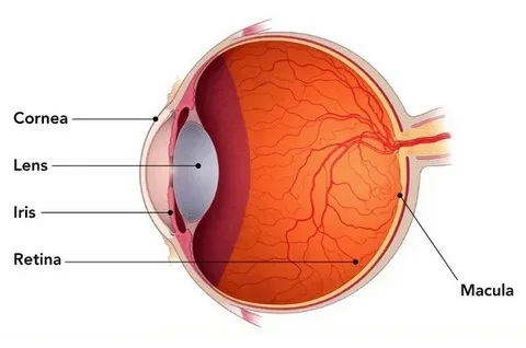
Retina is the light sensitive layer at the back of the eye. It receives the rays of light and sends it to the brain via the optic nerve for interpretation.
The retina has 10 layers and has blood vessels running through it.Macula is the central and the most sensitive area of the retina.
About Us
Age Related Macular Degeneration(ARMD) -
Macular degeneration
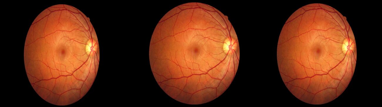
Macular degeneration is the deterioration of the macula (the central area of the reti-na responsible for detail vision). Central vision becomes blurred and distorted, and results in lost detail vision, difficulty in reading, increased need for light and reduced colour vision. There are two types of this disease: dry and wet. Dry is more common (90%). Due to blood vessel growth in or under the retina, the wet type results in severe vision loss, which occurs more rapidly than in cases of dry macular degeneration.
Although Macular Degeneration can occur during middle age, the risk increases, as a person gets older. Results of a large study show that people in their 50s have about a two percent chance of getting this problem. This risk rises to nearly 30 percent in those over age 75.
Certain studies have shown that it ranks second after diabetic retinopathy as the leading cause of blindness in the 45 to 64 year age group. Besides age, other risk factors include Neither dry nor wet AMD causes any pain. The most common symptom of dry AMD is slightly blurred vision. You may need more light for reading and other tasks. Also, you may find it hard to recognize faces until you are very close to them.
People with the dry type in one eye often do not notice any changes in their vision. With one eye seeing clearly, they can still drive, read, and see fine details. Some people may notice changes in their vision only if the condition affects both of their eyes
An early symptom of the wet type is that straight lines appear wavy.
Macular degeneration is the deterioration of the macula (the central area of the reti-na responsible for detail vision). Central vision becomes blurred and distorted, and results in lost detail vision, difficulty in reading, increased need for light and reduced colour vision. There are two types of this disease: dry and wet. Dry is more common (90%). Due to blood vessel growth in or under the retina, the wet type results in severe vision loss, which occurs more rapidly than in cases of dry macular degeneration.
Although Macular Degeneration can occur during middle age, the risk increases, as a person gets older. Results of a large study show that people in their 50s have about a two percent chance of getting this problem. This risk rises to nearly 30 percent in those over age 75.
Certain studies have shown that it ranks second after diabetic retinopathy as the leading cause of blindness in the 45 to 64 year age group. Besides age, other risk factors include Neither dry nor wet AMD causes any pain. The most common symptom of dry AMD is slightly blurred vision. You may need more light for reading and other tasks. Also, you may find it hard to recognize faces until you are very close to them.
People with the dry type in one eye often do not notice any changes in their vision. With one eye seeing clearly, they can still drive, read, and see fine details. Some people may notice changes in their vision only if the condition affects both of their eyes
Why Choose CEHC Retina Clinic in Chennai?
- Experienced Retina Specialist in Chennai
- Advanced OCT & OCT Angiography
- Digital Fundus Photography
- Fluorescein Angiography (FFA)
- Retina Laser Treatment
- Intravitreal Eye Injections
- Vitreo-Retinal Disease Management
- Personalized Treatment Plans
- Conveniently located in Thoraipakkam, Chennai
Meet Our Retina Specialist
Dr. Ashok Rangarajan is an experienced ophthalmologist with expertise in:
- Medical Retina
- Diabetic Retinopathy
- Macular Degeneration
- Retinal Vein Occlusion
- Retinal Laser Treatment
- Vitreo-Retinal Diseases
Frequently Asked Questions (FAQ)
The Retina Clinic diagnoses and treats a wide range of retinal diseases, including diabetic retinopathy, retinal vein occlusions, age-related macular degeneration (AMD), macular disorders, retinal vascular diseases, and other conditions affecting the back of the eye. Advanced diagnostic imaging and evidence-based treatments are used for comprehensive care.
Symptoms such as sudden blurred vision, flashes of light, floaters, distorted vision, dark spots, or loss of central or peripheral vision may indicate a retinal condition. If you experience any of these symptoms, seek an eye examination promptly.
Diabetic retinopathy is diagnosed through a detailed retinal examination and imaging tests such as fundus photography and OCT/OCTA. Depending on the severity, treatment may include retinal laser therapy, intravitreal injections, or surgery to preserve vision.
Optical Coherence Tomography Angiography (OCTA) is a non-invasive imaging technique that provides detailed views of retinal blood vessels without dye injection. It helps detect and monitor conditions such as diabetic retinopathy and macular degeneration with high precision.
While AMD cannot always be cured, early diagnosis and appropriate treatment can help slow disease progression and preserve vision. Management depends on whether the condition is the dry or wet form and may include medications, injections, and regular monitoring.
Intravitreal injections deliver medication directly into the eye to treat retinal diseases such as wet AMD and diabetic macular edema. They are performed under sterile conditions and are widely used as a safe and effective treatment option.
Most retinal laser procedures are performed on an outpatient basis and do not require an overnight hospital stay. Patients can usually return home the same day after receiving post-procedure instructions.
Yes. Diabetic retinopathy can develop without noticeable symptoms in its early stages. People with diabetes should undergo regular retinal examinations to detect changes early and reduce the risk of vision loss.
Individuals with diabetes or hypertension should follow their ophthalmologist’s recommendations for regular retinal screening. The frequency depends on their eye findings and overall health, but periodic examinations are essential for early detection of retinal damage.
A retina evaluation may include a dilated eye examination, fundus photography, OCT or OCTA scans, fluorescein angiography in selected cases, and other imaging tests to accurately diagnose and monitor retinal conditions.
Some retinal diseases can cause irreversible vision damage if left untreated. Early diagnosis and timely intervention significantly improve the chances of preserving vision and preventing complications.
You can schedule a consultation with the Retina Clinic for a comprehensive retinal evaluation, advanced diagnostic testing, and personalized treatment planning based on your specific eye condition.
If you are looking for an experienced retina specialist in Chennai, Dr. Ashok Rangarajan at CEHC (Dr. Ashok Eye Hospital) is a trusted ophthalmic surgeon with over 30 years of experience in retina, cataract, glaucoma, and comprehensive eye care. Patients from Chennai and surrounding areas visit CEHC for advanced retinal evaluation and personalized treatment plans.
You can receive comprehensive retina treatment at CEHC – Dr. Ashok Eye Hospital in Thoraipakkam, Chennai. The hospital offers advanced retinal diagnostics, OCT scans, fundus imaging, laser treatment, intravitreal injections, and management of complex retinal diseases under the care of experienced specialists.
CEHC treats a wide range of retinal disorders, including diabetic retinopathy, age-related macular degeneration (AMD), retinal tears, retinal detachment, macular edema, retinal vein occlusion, and other vision-threatening retinal diseases using modern diagnostic and treatment technologies
You should consult a retina specialist immediately if you experience sudden vision loss, flashes of light, floaters, distorted vision, blurred central vision, or have diabetes with changes in your eyesight. Early diagnosis can help prevent permanent vision loss.
Yes. Dr. Ashok Eye Hospital - Unit of Centre for Eye and Health Care (CEHC) offers comprehensive diabetic retina screening using advanced retinal imaging to detect diabetic retinopathy at an early stage. Regular eye examinations are recommended for every diabetic patient to protect their vision.
Yes, many hospitals assist patients by providing the necessary medical records, estimates, invoices, and supporting documents required for insurance reimbursement or claim processing.